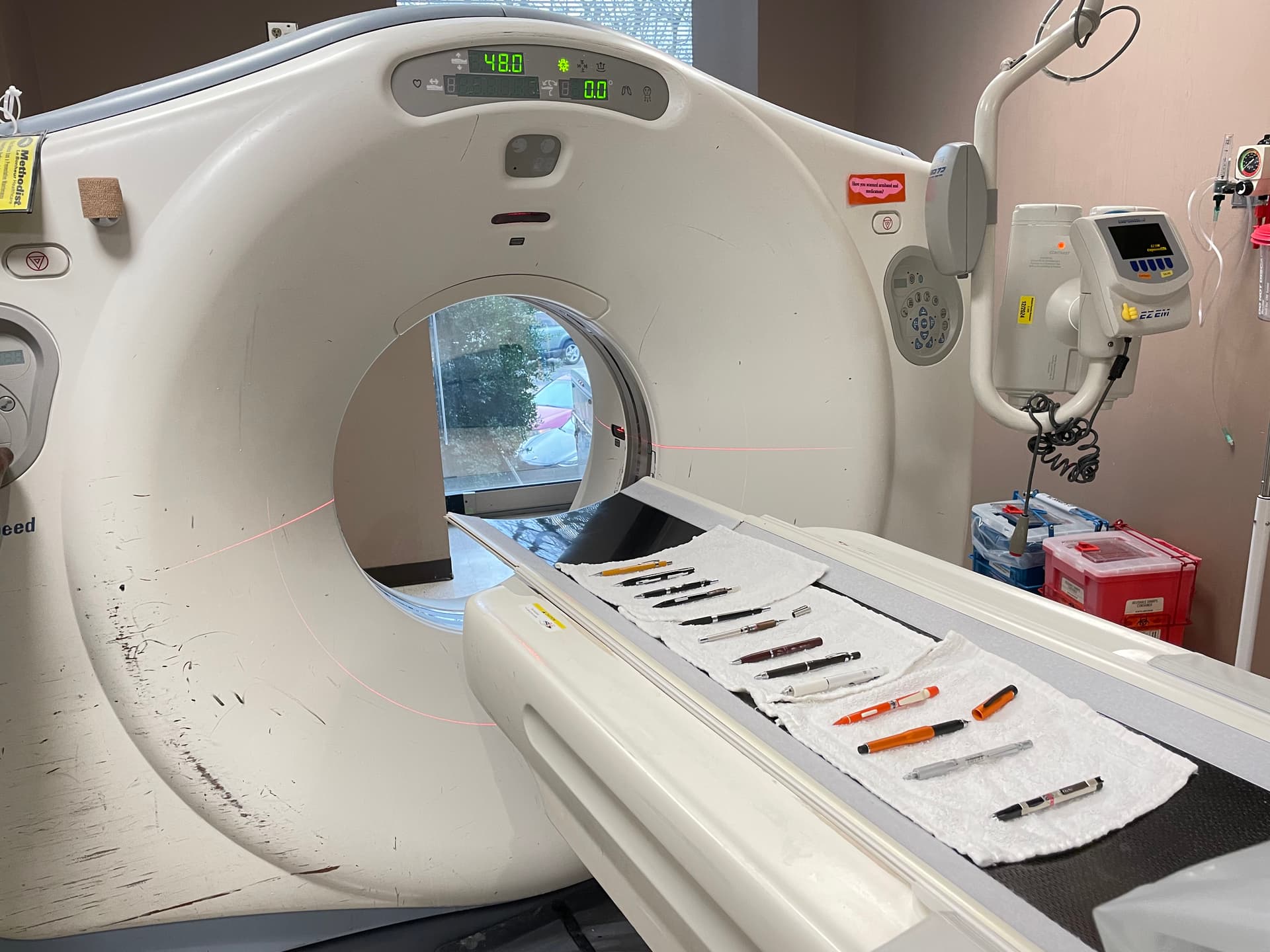
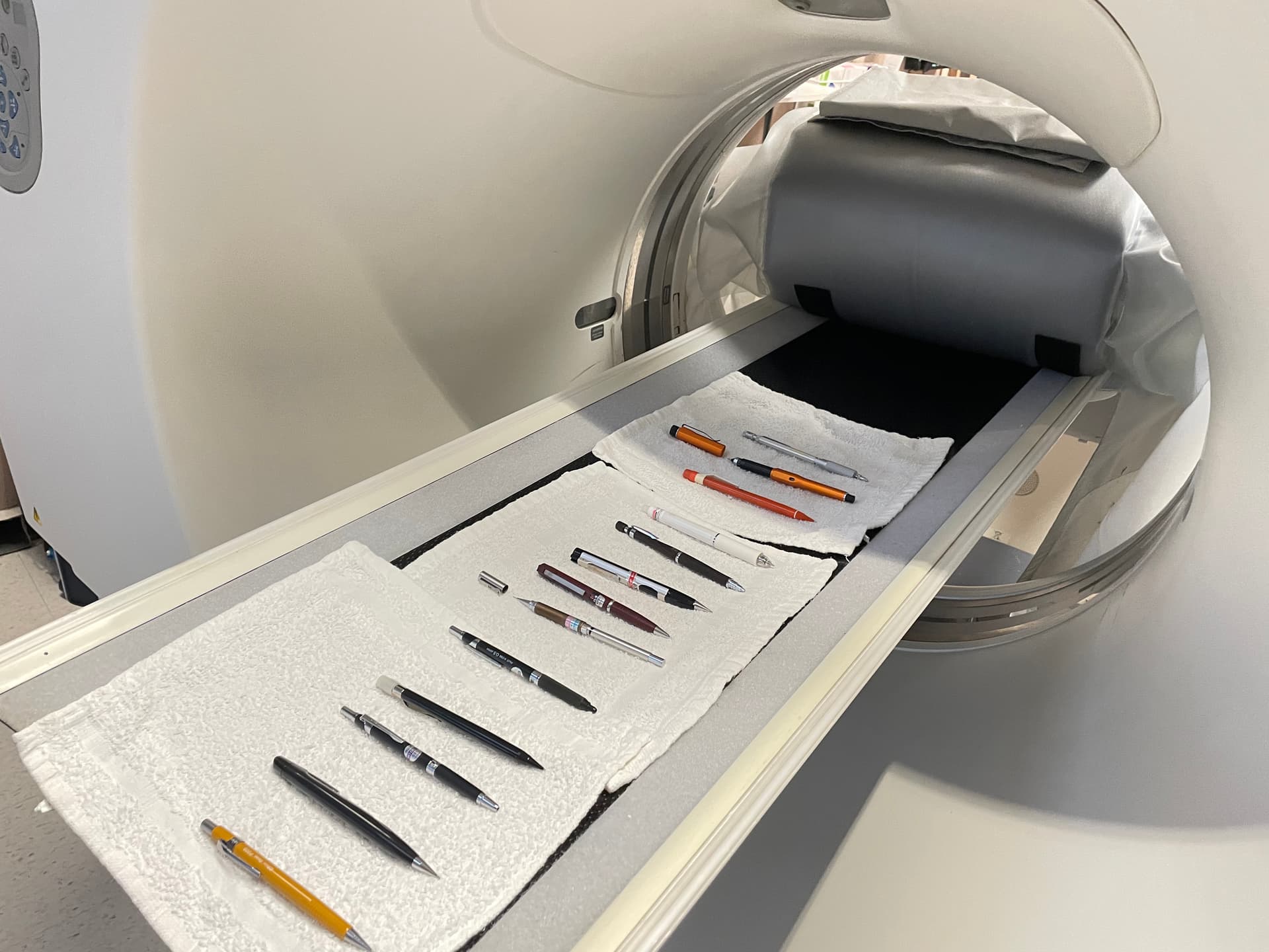
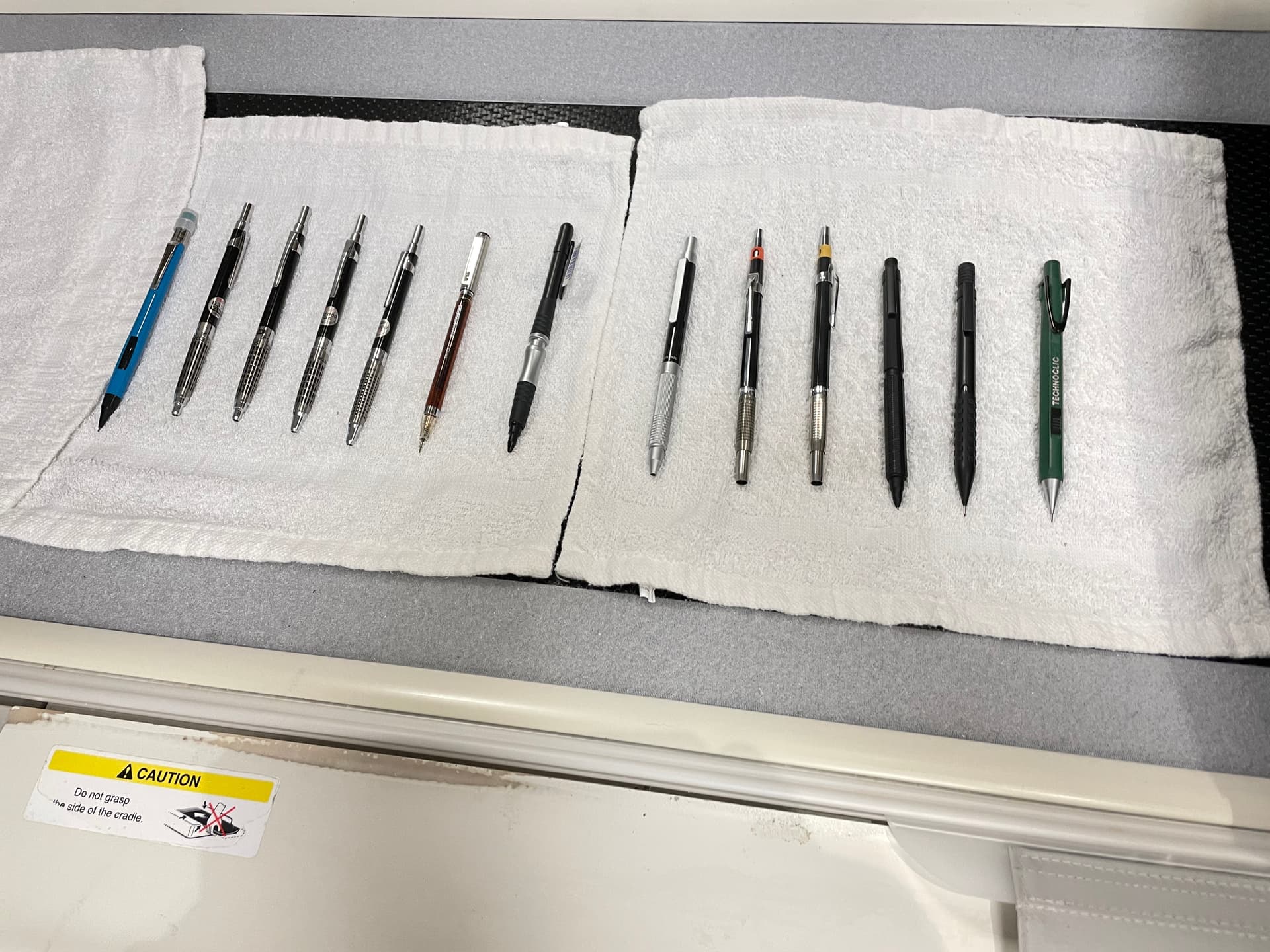
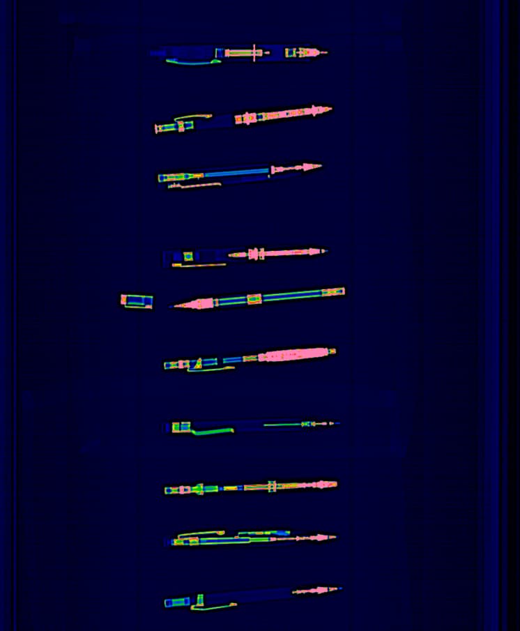
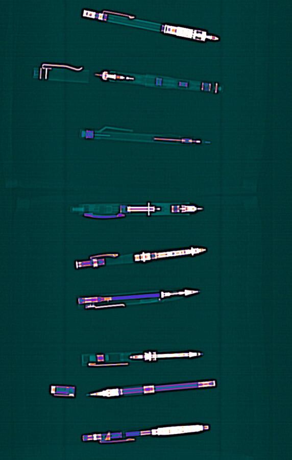
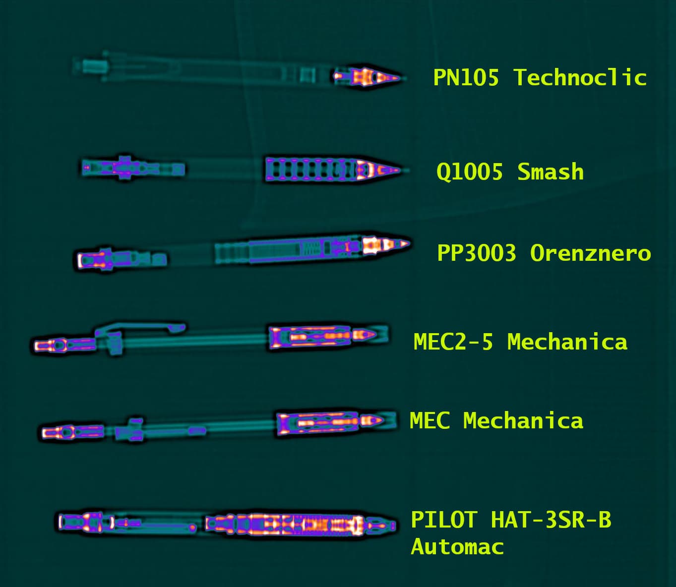
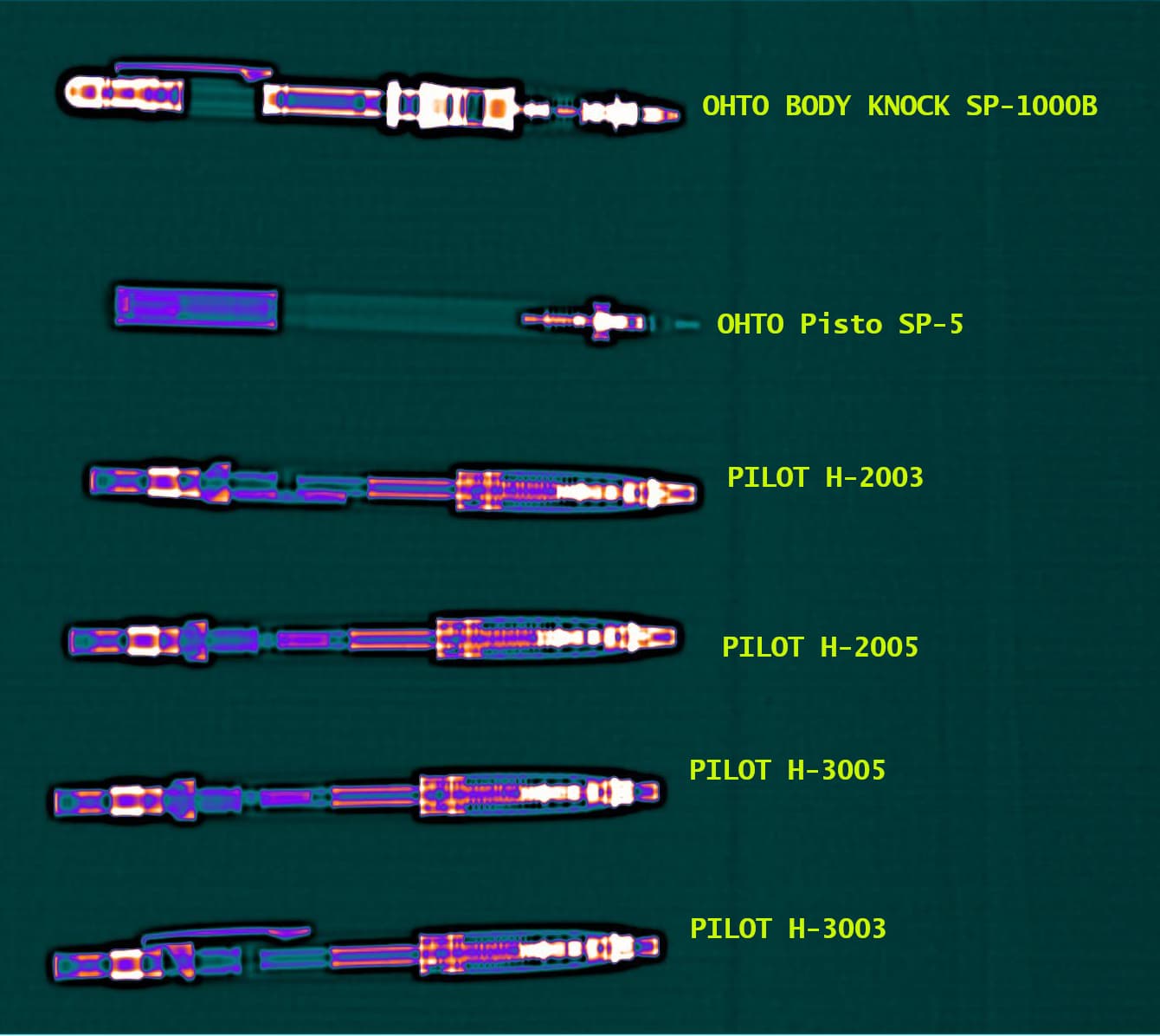
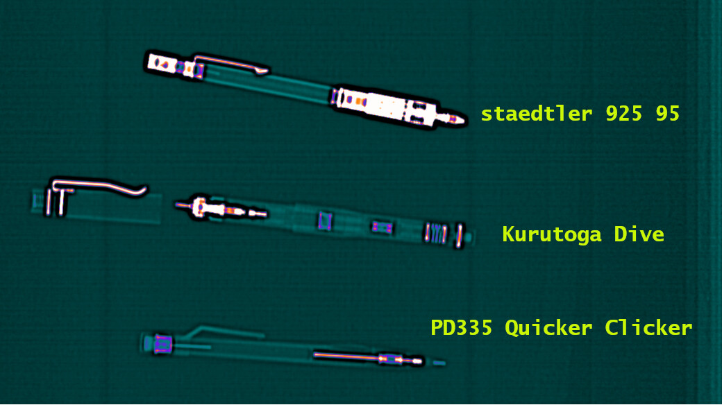
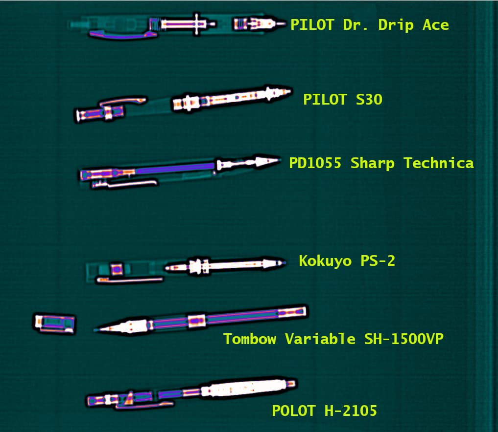
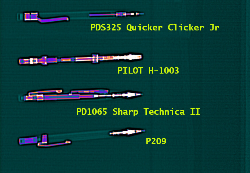
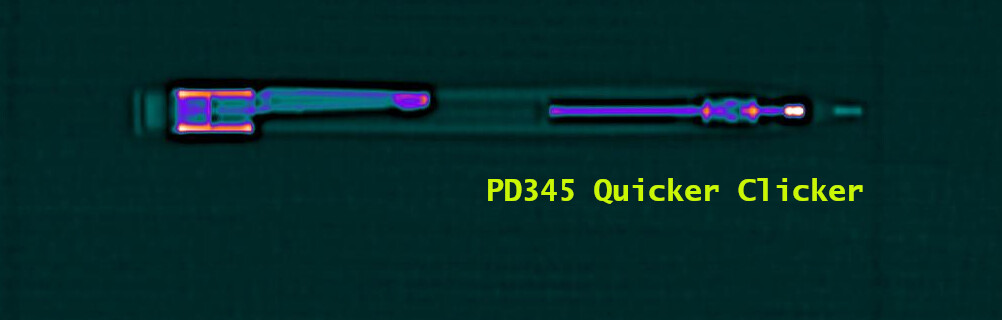
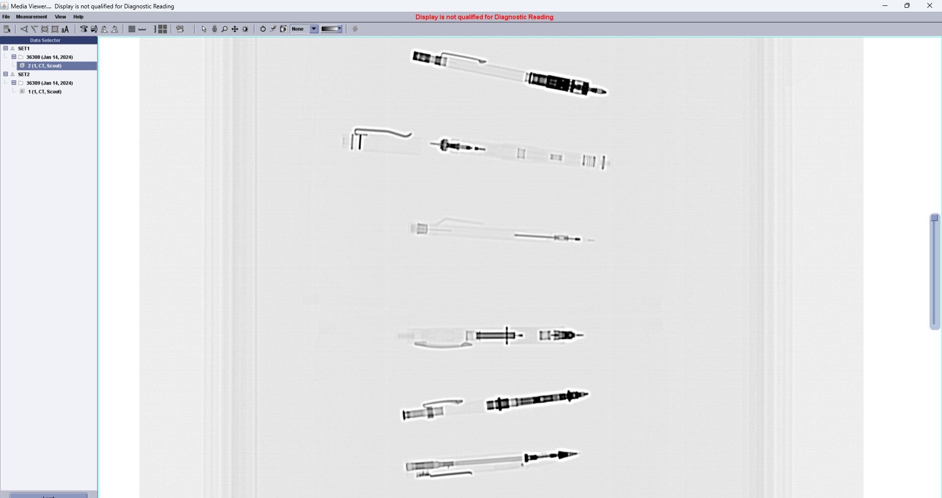
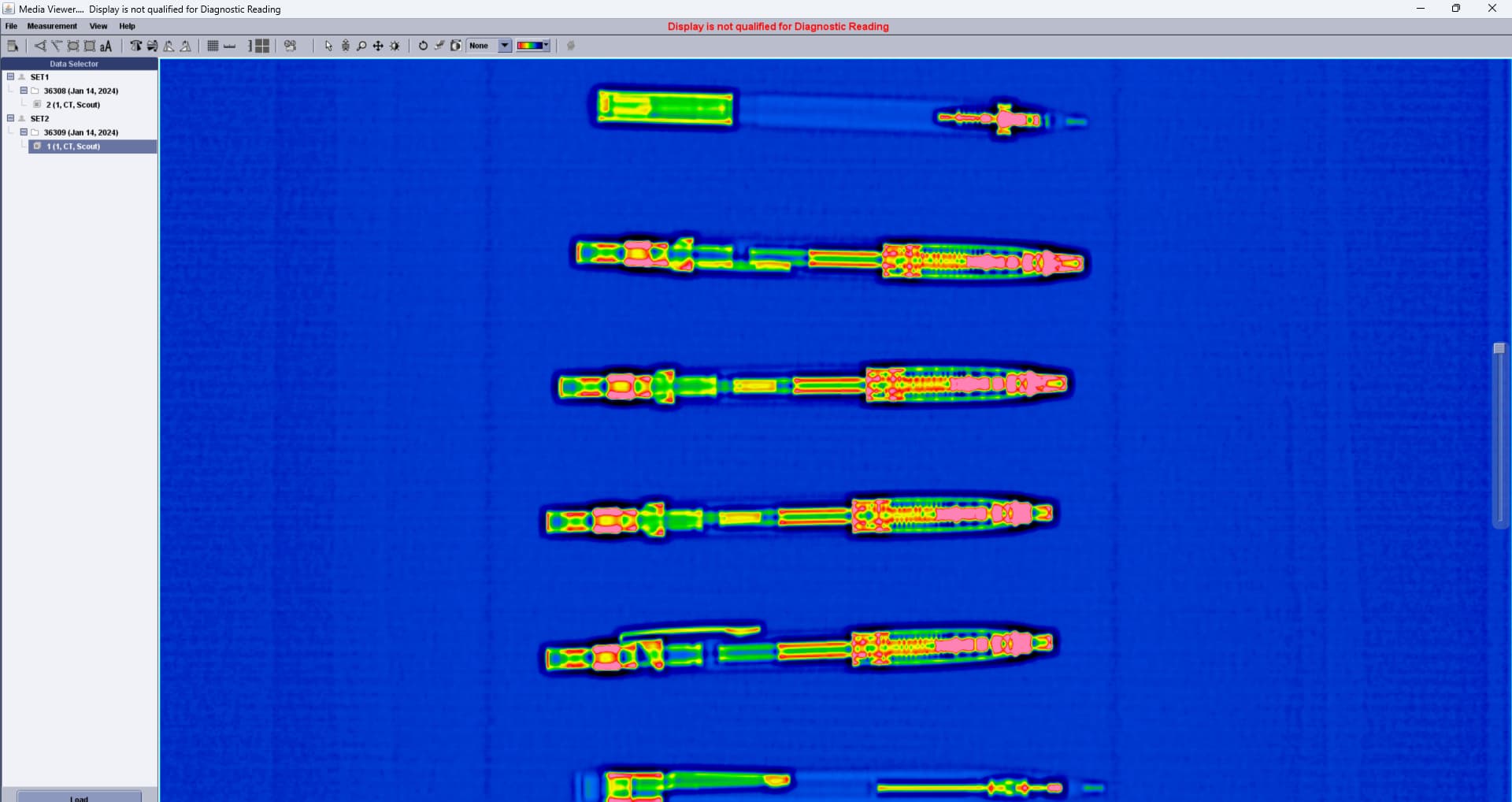
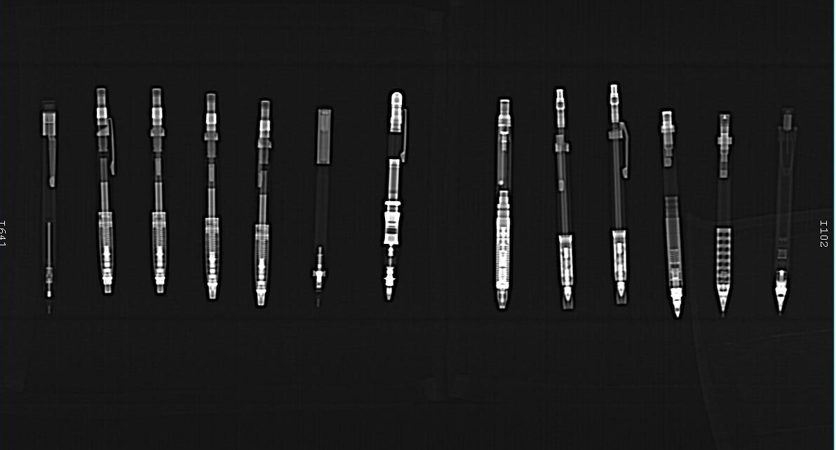
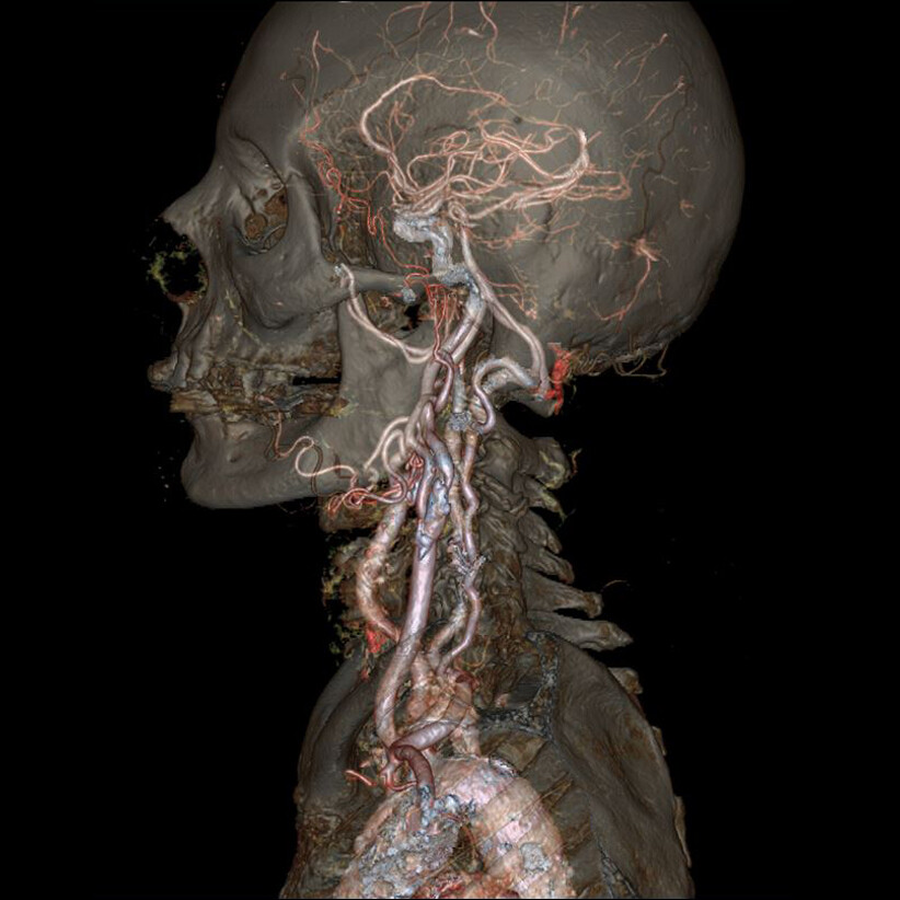
Yesterday I was able to meet up with a buddy of mine to try to get some CT scans of a few mechanical pencils. This particular CT scanner was pretty old, and only a 64 slice system, but it did have the latest software available to that model. Anyway, this thing is like magic when imaging the human body. The surgeon is able to select the particular part of the patient’s physiology that is being imaged, and separate it from the rest, and even view it in 3D (spin it around, etc). The first image below is an example of how this device works with soft tissues. It’s amazing what this system can do. It’s mostly image manipulation w/ software post scan. But to he honest this scanner is old as crap. Our newest 256 slice scanners make this one look amateurish.
Anyway, we tried for a few hours, but this system just isn’t made to look at mechanical pencils. I am happy with the results we got, but they are a bit washed out, over saturated, etc. We couldn’t do a “normal” scan – these are actually what they call “scout” scans – low intensity, low resolution scans. They are a pre-scan that they perform just to be sure everything is “OK” before the real scan.
I am working on a rig of sorts made from clear acrylic tube and rods that will allow us to capture a Kurutoga Dive under X-Ray Fluoroscopy. This will enable us to grab a video of what goes on inside while it is writing, spinning and advancing lead, etc.
Getting access to the 256 slice scanner could possibly happen when it is taken down for its next routine maintenance schedule. But the higher end imaging devices are in very high demand, so once it’s certified, if there are patients waiting, I’ll have to wait another 3 months for the next PM.
We may have better luck using a PET scanner, but that also is in very high demand.
I really want to get some videos of the Dive working. I’ll try to make that happen sometime soon if I can.